The Worlds Within Our Body
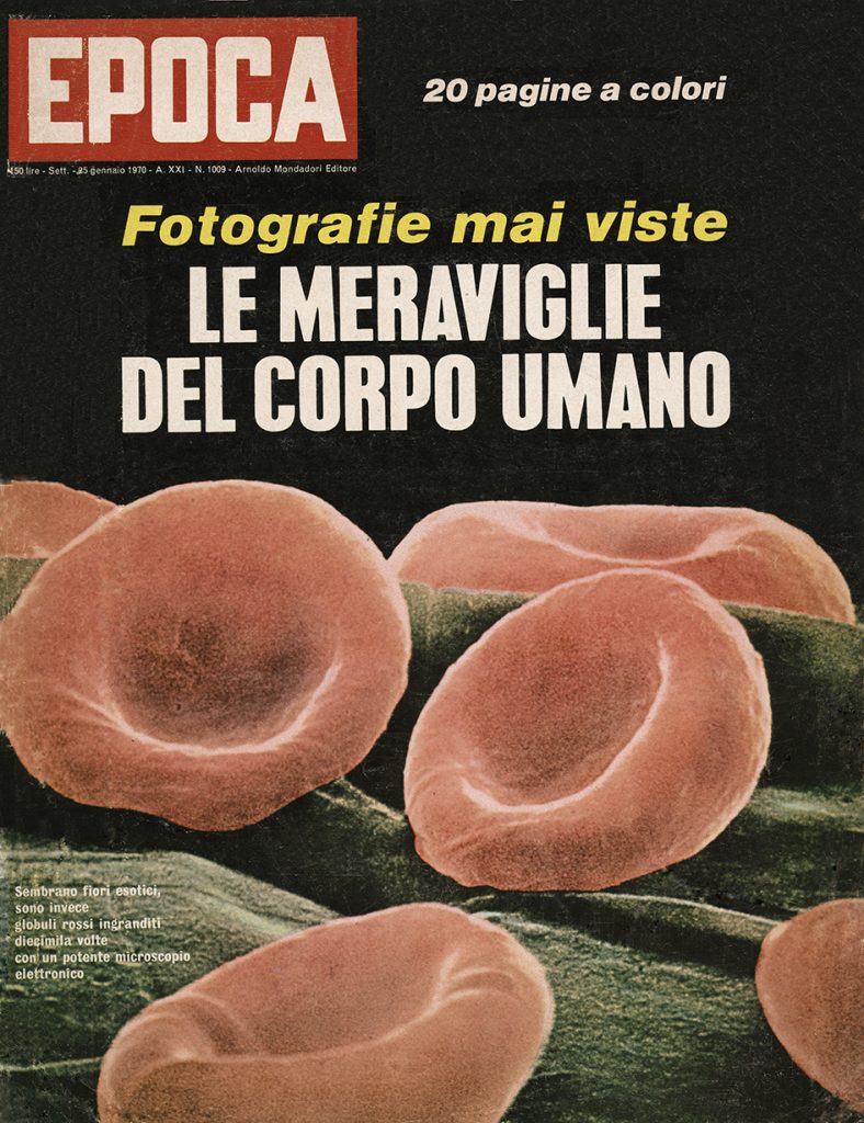
I Life Magazine, den 9 januari 1970, visar Lennart sina första bilder tagna med hjälp av svepelektronmikroskop. Det är också de första SEM bilderna som Gillis Häägg färgat.
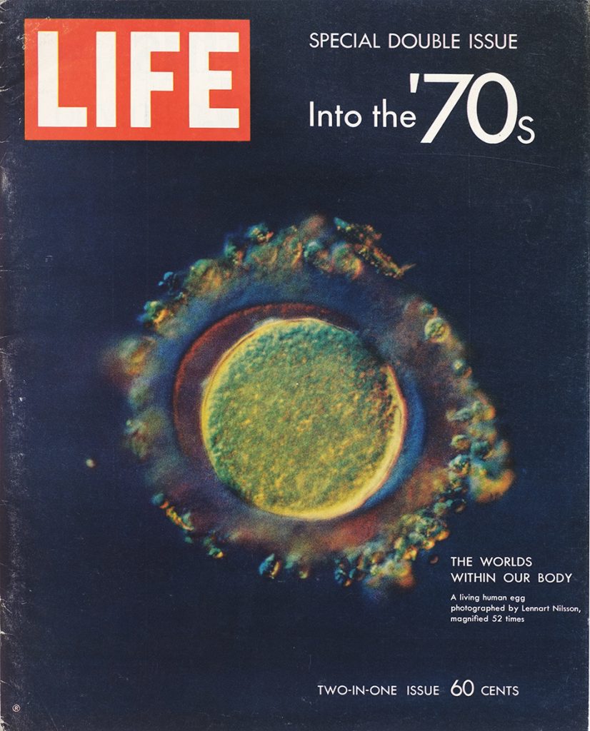
Along the way he assisted scientists with their own research, participated in scientific papers – and pioneered a variety of photographic techniques
Life Magazine, 9 January 1970
-
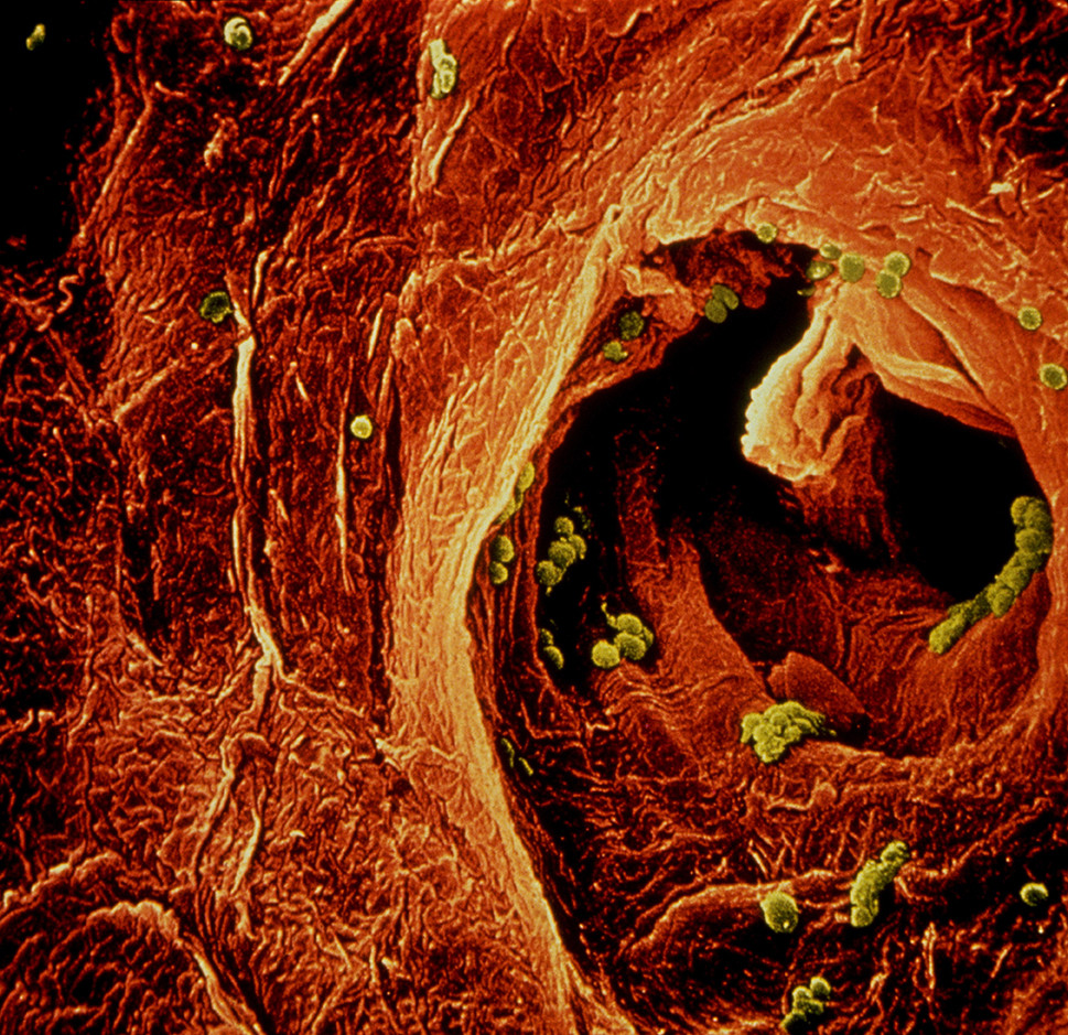
En öppning i fingertoppen där man ser svett komma ut. De gröna "bollarna" är bakterier, 1970. ©Lennart Nilsson/SPL -
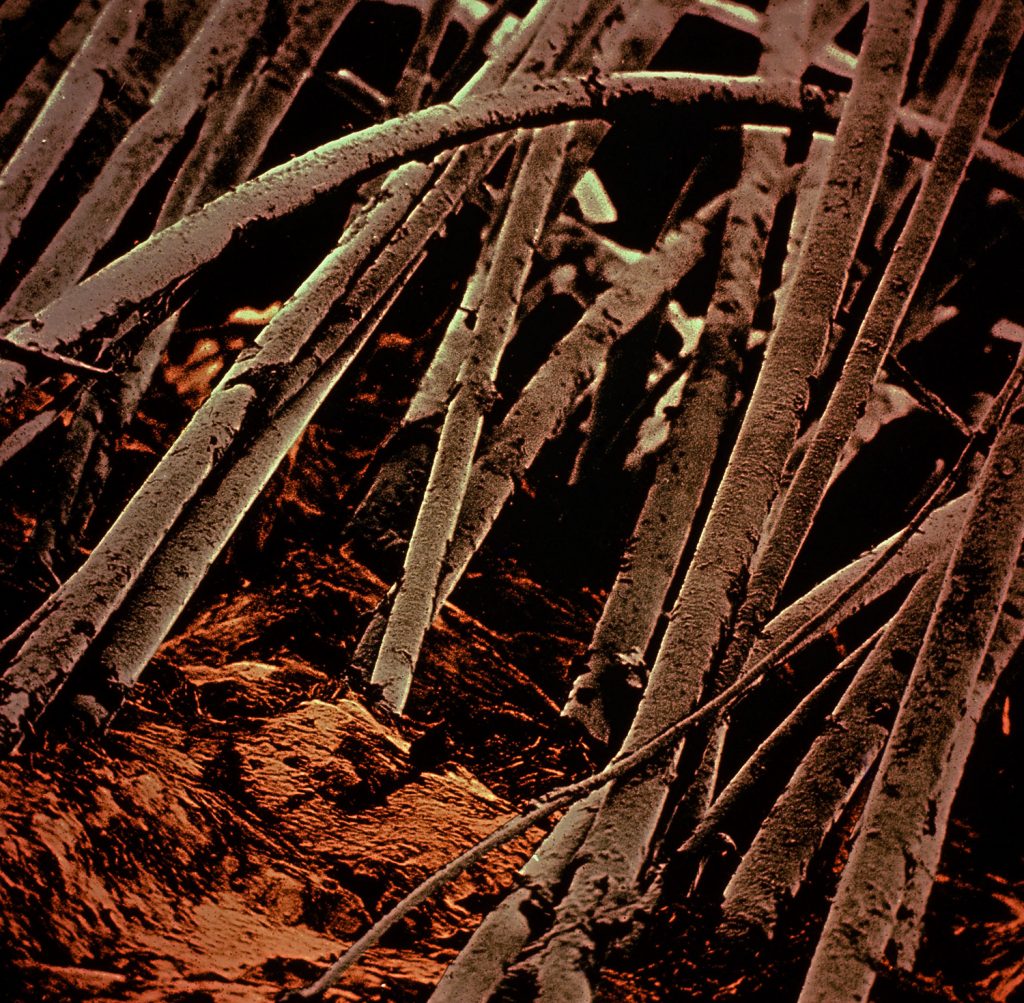
Hår på huvudet. Första bilderna med svepelektronmikroskopi 1969. Färglagd av Gillis Häägg samma år. ©Lennart Nilsson/SPL -
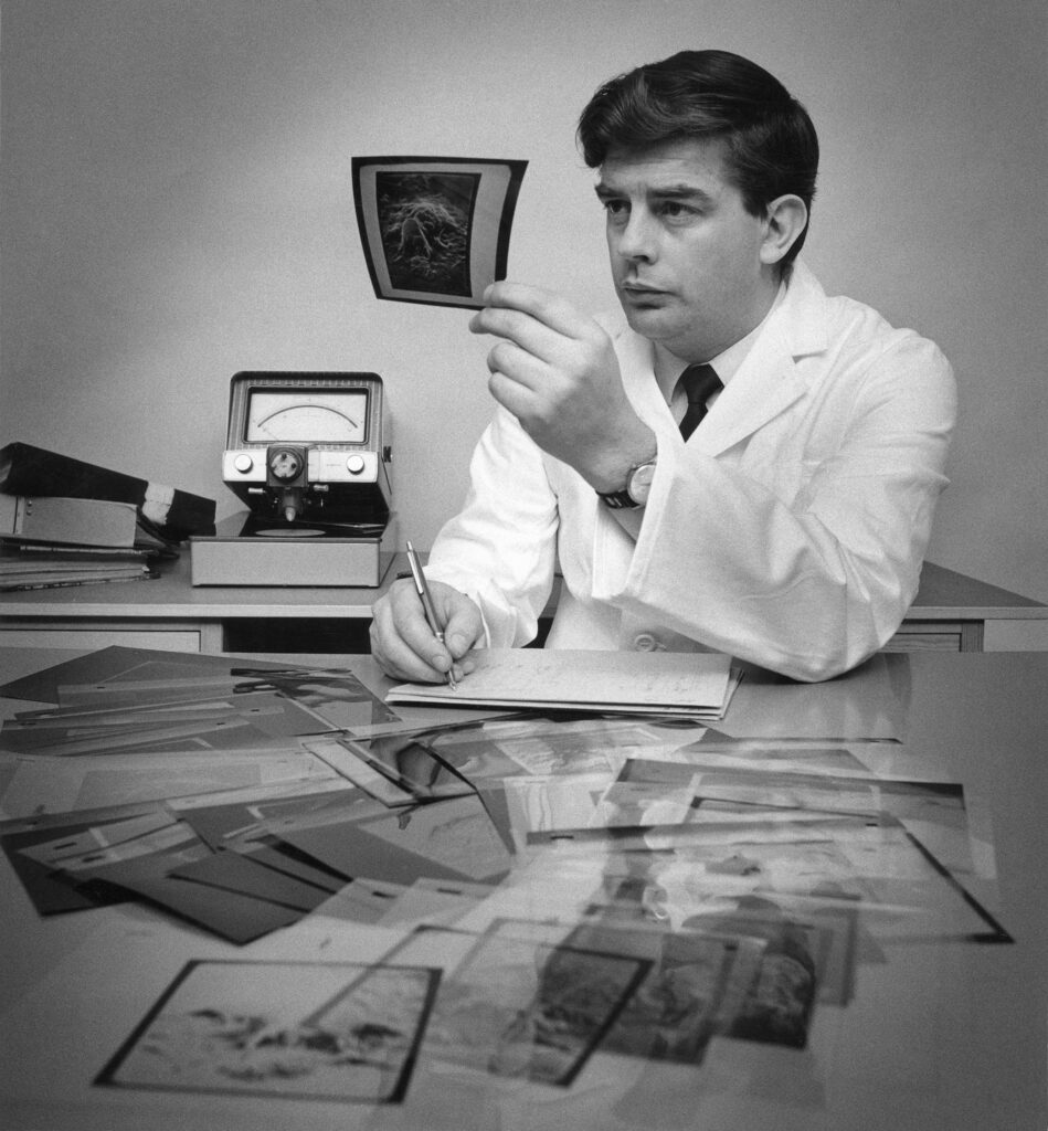
Gillis Hääg, 1970 -
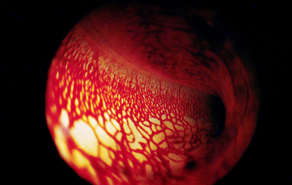
I trumhåletrappan i innerörat, 1970. ©Lennart Nilsson/SPL -
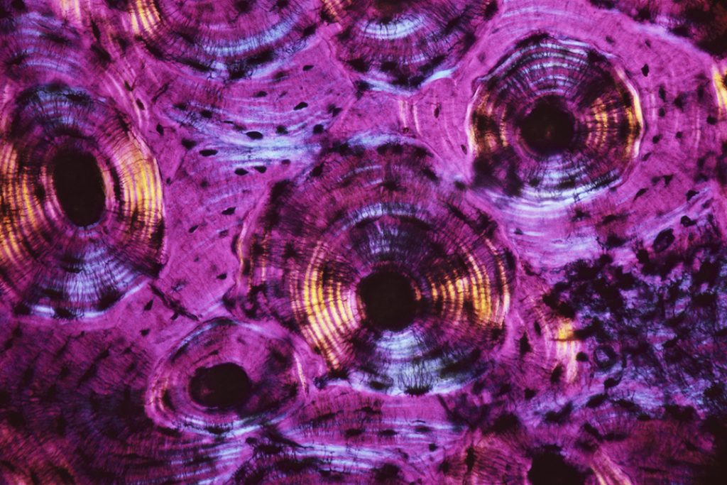
Benvävnad i mikroskopiskt tvärsnitt, 1970. ©Lennart Nilsson/SPL -
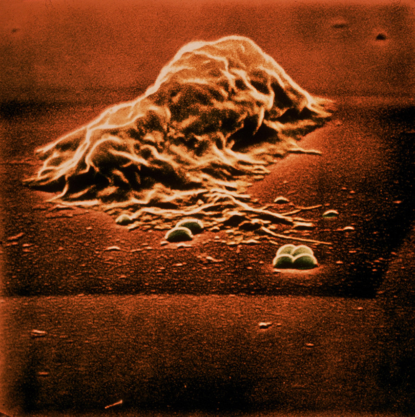
Kampen mellan vit blodkropp (lymfocyt) och bakterier, 1969 ©Lennart Nilsson/SPL -
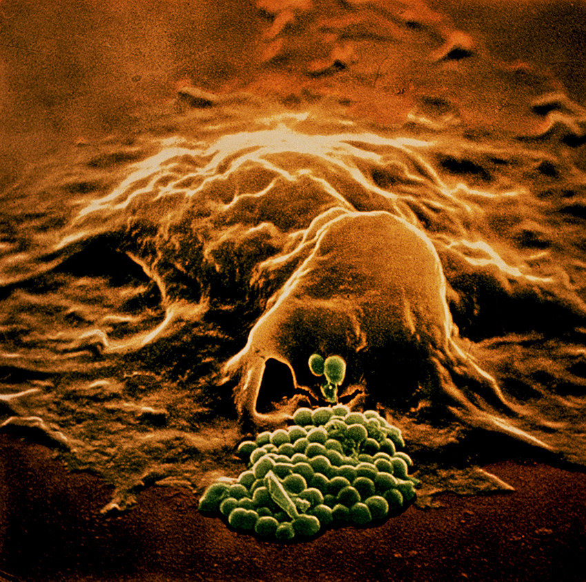
Kampen mellan vit blodkropp (lymfocyt) och bakterier, 1969. ©Lennart Nilssson/SPL -
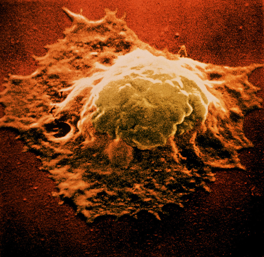
Kampen mellan vit blodkropp (lymfocyt) och bakterier, 1969. ©Lennart Nilsson/SPL -
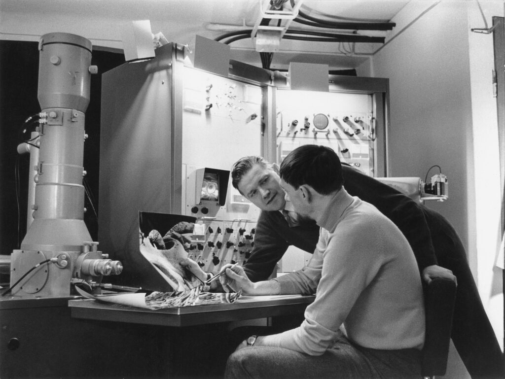
Lennart Nilsson och Göran Alsterborg på Analytica i Stockholm 1969
-

En öppning i fingertoppen där man ser svett komma ut. De gröna "bollarna" är bakterier, 1970. ©Lennart Nilsson/SPL -

Hår på huvudet. Första bilderna med svepelektronmikroskopi 1969. Färglagd av Gillis Häägg samma år. ©Lennart Nilsson/SPL -

Gillis Hääg, 1970 -

I trumhåletrappan i innerörat, 1970. ©Lennart Nilsson/SPL -

Benvävnad i mikroskopiskt tvärsnitt, 1970. ©Lennart Nilsson/SPL -
Kampen mellan vit blodkropp (lymfocyt) och bakterier, 1969 ©Lennart Nilsson/SPL 
-

Kampen mellan vit blodkropp (lymfocyt) och bakterier, 1969. ©Lennart Nilssson/SPL -

Kampen mellan vit blodkropp (lymfocyt) och bakterier, 1969. ©Lennart Nilsson/SPL -

Lennart Nilsson och Göran Alsterborg på Analytica i Stockholm 1969
“This extraordinary series of electron microscope pictures shows a large white cell known as a macrophage actually consuming dangerous invaders, staphylococci bacteria. To make these pictures, (the last three images), Photographer Nilsson scraped a few white cells from his own throat, obtained bacteria from Swedish bacteriologists and put them together under a standard light microscope – with a supply of penicillin on hand in case of accidental infection. As each white cell reached a particular stage in its attack on the bacteria, he stopped the process with a fixative chemical and began to job of preparing the specimen for the electron microscope – washing, cleaning and coating it with a delicate gold film designed to reflect electrons and, ultimately, produce the cell’s image on a phosphorescent screen. Nilsson then photographed the screen and, with the help of fellow researchers, added color that matched the original cells as closely as possible. The final sequence provides a remarkable record of the kind of crucial battle that goes on constantly – and invisibly – in our bodies.”
Se reportaget i Life Magazine
